Anatomy of the Body
Why is the VMO Muscle so Important?
As a Pilates instructor it is important to understand how Pilates can help strengthen specific muscles. The Vastus Medialis Oblique (VMO) is essential for knee stability and keeping the Patella (kneecap) aligned when the leg extends. The VMO is key to maintaining strong knees and avoiding knee pain, making it a fantastic muscle to target when instructing Pilates.
WHAT IS THE VMO?
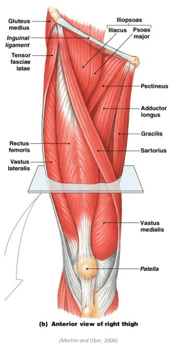
The Vastus Medialis Oblique (VMO) is one half of the Vastus Medialis muscle, sitting just above the Patella and along the inside of the thigh (Tenan et al., 2013). Along with the Vastus Intermedius, Vastus Lateralis, and Rectus Femoris – the Vastus Medalis is one of the four Quadricep muscles. While all four Quadricep muscles work to extend the knee, the VMO also helps to stabilise the knee (Peng et al, 2017). When the knee straightens and bends, the Patella moves up and down (Tenan et al., 2013). As this movement happens, the VMO helps keep the Patella on track when moving back into the Trochlear Groove (kneecap groove). This function is incredibly important as it helps to avoid injuries, such as knee dislocation.
Let’s revise some VMO anatomy!
Origin: Shaft of the Femur
Insertion: Patella
Action:
- Extends and stabilises the knee
- Draws the Patella medially
IMPORTANCE OF THE VMO?
The VMO is essential in helping the knee track correctly and maintaining stability, which is needed for everyday activities, such as walking and participating in exercise. When the knee is functioning correctly, there should be smooth tracking of the Patella when the knee extends and contracts.
However, if there is fluid, swelling, or an injury around the knee, it can stop the VMO from working correctly, causing the VMO to become weak. If the VMO is weak or fatigued, the Patella can mistrack which results in knee pain. This is because the Patella is not moving smoothly back into the Trochlear Groove.
Having a strong VMO can not only help prevent injuries, but can also assist in the rehabilitation of knee injuries. But how do you strengthen the VMO?
BEST PILATES EXERCISES FOR DEVELOPING VMO STRENGTH
To help strengthen the VMO muscle, it is important to program Pilates exercises that help activate the VMO as well as the other Quadricep muscles (Peng et al, 2017).
FOOTWORK 1 (PARALLEL)
INHALE: T-Zone and squeeze the buttocks
EXHALE: press through the heels to press
the carriage out, straightening the knees
INHALE: bend the knees, controlling the
carriage back in
Footwork (Parallel) is a great exercise to strengthen the VMO and is usually better tolerated than Footwork (Internal Rotation) or Footwork (External Rotation) if a client is experiencing knee pain.
FOOTWORK 4 (INTERNAL ROTATION)
INHALE: T-Zone and squeeze the buttocks
EXHALE: press through the heels to press
the carriage out, straightening the knees
INHALE: bend the knees, controlling the
carriage back in
The internal rotation used in Footwork 4 can be more helpful in activating the VMO than the other footwork positions. Clients can also squeeze a cushion between their knees while performing this exercise to bias the VMO more. This occurs because squeezing a cushion keeps the legs internally rotated the entire time.
SINGLE LEG STRETCH 1 (TABLETOP)

INHALE: T-Zone and squeeze the buttocks
EXHALE: press through the heel to press
the carriage away, straightening the knee
INHALE: bend the knee, controlling the
carriage back in
This exercise is a great option because it works unilaterally, which means that you can focus more on one particular leg if there is an injury and also ensures that one leg is not overcompensating for the other.
BICYCLE LEGS + BAND
INHALE: T-Zone
EXHALE: curl up and draw the ribs to the hips, lifting the head and shoulders, hold this position
INHALE: T-Zone
EXHALE: press into the band, extending the leg out, straightening the knee
INHALE: return the leg back in
Continue on the same side
Make sure to cue clients to activate the VMO (drawing the kneecap upward and inward as the leg extends)
SQUATS + CIRCLE (BETWEEN THIGHS)
INHALE: squat down bending the knees, sticking the bottom out
EXHALE: squeeze the buttocks to stand back up
Using the circle between the thighs also helps the T-Zone and VMO muscles to work better by placing the legs in the correct alignment. When the circle is used in between the thighs, ensure that the circle is placed above the knees (not over the knee joint itself) and it is evenly spaced between both knees so that the handles sit flat against the inner thighs.
WALL SQUATS + BALL
INHALE: squat down bending the knees, sticking the bottom out”
EXHALE: squeeze the buttocks to stand back up
Add a circle between the knees or a band around the thighs to help with the VMO and Gluteal activation and assist with correct knee alignment throughout the exercise
OTHER TIPS TO AID VMO ACTIVATION
- Progress to gradual strengthening of the VMO and buttocks, making sure there is never any pain
- To help the VMO to activate, press the heel into the bar and focus on squeezing the Quadriceps just above the knee on the medial side. Getting the clients to palpate here and feel if the muscle is activating can helpful
- Make sure that the feet are in the correct position (ie. not rolling in and not twisting out) as this can put strain on the knee
PALPATING THE VMO
Check out the below vidoes for more information on palpating the VMO
REFERENCES
Martini, F. and Ober, W. (2006). Fundamentals of Anatomy & Physiology. San Francisco, CA: Pearson Benjamin Cummings.
Tenan, M. S., Peng, Y.-L., Hackney, A. C., & Griffin, L. (2013). Menstrual Cycle Mediates Vastus Medialis and Vastus Medialis Oblique Muscle Activity. Medicine and Science in Sports and Exercise, 45(11), 2151–2157. https://doi.org/10.1249/MSS.0b013e318299a69d
Peng, Y.-L., Tenan, M. S., & Griffin, L. (2018). Hip position and sex differences in motor unit firing patterns of the vastus medialis and vastus medialis oblique in healthy individuals. Journal of Applied Physiology (1985), 124(6), 1438–1446. https://doi.org/10.1152/japplphysiol.00702.2017